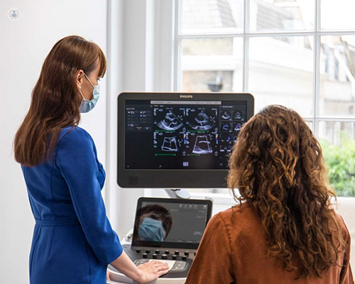
Transoesophageal echocardiography (TOE): a comprehensive guide
Written in association with:Transoesophageal echocardiography (TOE) is a specialised, sophisticated ultrasound that provides an exceptionally detailed look at your heart and its adjacent structures. Imagine a high-tech camera that can see through the usual barriers, giving your doctor an insider's view of your heart's inner workings. That's what TOE offers, making it a game-changer in heart care.
Leading consultant cardiologist Dr Gosia Wamil answers your questions about transoesophageal echocardiography (TOE) including when it may be necessary, what it involves and the recovery process.

How has transoesophageal echocardiography evolved over time?
Transoesophageal echocardiography (TOE) has undergone remarkable advancements since its inception in the 1970s. Originally experimented with as a diagnostic tool, TOE has evolved into a vital component of cardiac imaging technology. Its distinguishing feature lies in the placement of the probe within the oesophagus, situated just behind the heart. This strategic positioning eliminates the obstacles typically encountered in conventional transthoracic echocardiography, such as interference from ribs or lung tissue.
Why may you need a TOE?
Transoesophageal echocardiography (TOE) may be necessary for several reasons in cardiac care. It's particularly useful if you have abnormal heart valve functions or if a standard echocardiogram (done from outside the chest) suggests there might be something more to explore.
It offers detailed imaging of the heart and surrounding structures, providing crucial information for diagnosing various heart conditions such as valve disorders, congenital heart defects, blood clots, or abnormal heart rhythms. TOE is particularly useful when traditional echocardiography techniques are limited due to factors like obesity, lung disease, or chest deformities. Additionally, it aids in guiding interventions like cardiac surgeries or procedures such as closure of atrial septal defects.
What happens during a TOE?
While the thought of a medical procedure might be daunting, knowing what to expect can ease some anxiety. Typically, a TOE takes about an hour, with some additional observation time afterwards. The procedure involves a small, flexible tube (about the size of a little finger) with an ultrasound device at its tip. This tube is gently guided down your throat while you're sedated, allowing you to be comfortable and relaxed.
Before the procedure, you'll be given a local anaesthetic in your throat to minimize any discomfort, and sedation helps you stay relaxed. During the TOE, you'll lie on your side, and the healthcare team will monitor your vital signs closely. The images obtained by the TOE are incredibly detailed, offering real-time insights into your heart's condition.
What is the recovery process like?
The recovery process for transoesophageal echocardiography (TOE) is typically swift and straightforward. After the procedure, patients are usually monitored for a short period to ensure there are no immediate complications. Post-TOE, you'll need a little time to let the anaesthesia wear off, especially before eating or drinking.
Patients may be advised to avoid eating or drinking for a short period after the procedure to allow the effects of the sedation to wear off fully. It's normal to have a minor sore throat afterwards, which can usually be soothed with lozenges or mild pain relievers. Remember, the sedation can temporarily impact your judgment, so having someone assist you with mobility is wise.
Overall, the recovery process for TOE is generally well-tolerated, and patients can typically return to their usual routine shortly after the procedure.
Safety and outcomes
Transoesophageal echocardiography (TOE) is generally safe but carries minimal risks, including throat discomfort, gag reflex, and mild sedation-related complications. In rare cases, there's a risk of oesophageal injury, aspiration, or adverse reactions to sedatives. Serious complications such as bleeding or infection are extremely rare.
However, these risks are outweighed by the significant diagnostic benefits TOE offers in assessing cardiac conditions, especially when conventional echocardiography is insufficient.
It's a critical tool that has transformed how cardiologists assess and manage heart conditions, often providing essential information that can influence the approach to treatment or surgery. This procedure offers a unique glimpse into your heart's structure and function, providing invaluable information that helps shape your care. With TOE, you and your healthcare team can navigate your heart health journey with detailed maps and plans, ensuring the best outcomes for your well-being.
If you require a TOE and would to book a consultation with Dr Wamil, do not hesitate to do so by visiting her Top Doctors profile today.


