Abnormal chest X-ray

What is an abnormal chest x-ray?
A chest X-ray is an imaging test that utilises low doses of radiation in short blasts to create images of the inside of a patient’s chest. In this way, doctors can examine the heart, lungs, bones, and blood vessels.
If the X-ray images show abnormalities, this means that there is something unusual on the image of the chest. This is usually indicative of a problem, and could be immediately obvious, such as a broken or fractured rib, or could simply be a shadow that needs further investigation.
Some doctors specialise in dealing with abnormalities in chest X-ray results, and may order follow-up tests to determine the cause.
Why does a doctor order a chest x-ray?
A doctor may order a chest x-ray if a patient is presenting with symptoms such as:
- persistent coughing
- difficulty breathing
- wheezing
- coughing up blood
- chest pain
- chest injury
- fever
- shortness of breath
Other reasons a chest x-ray may be ordered is to reveal:
- fractures
- heart-related problems
- problems with blood vessels
- changes after having an operation
- lung conditions
- outline and size of the heart
- position of pacemaker, defibrillator, or catheter
What causes an abnormal chest X-ray?
An abnormal chest X-ray can be caused by a number of conditions.
Lungs:
- pneumonia (unusual white or hazy shadow on the normally dark lungs on the X-ray can indicate this)
- abscesses
- pulmonary oedema (fluid build-up in the lungs)
- lung cancer and other masses in the lungs
- cavities in the lungs or cavitary lesions (caused by diseases like tuberculosis and sarcoidosis)
- bronchitis
- asthma
Heart:
- an enlarged heart (cardiomegaly), which can, in turn, be caused by various conditions, such as hypertension or coronary artery disease.
- heart failure
- fluid around the heart
Bones:
- broken ribs
- broken or fractured vertebrae
- dislocated shoulder
Other:
- pleural effusion (fluid build-up between the lungs and chest wall)
- pneumothorax (build-up of air between the lungs and chest wall)
- hiatal hernia
- aortic aneurysm
- cysts
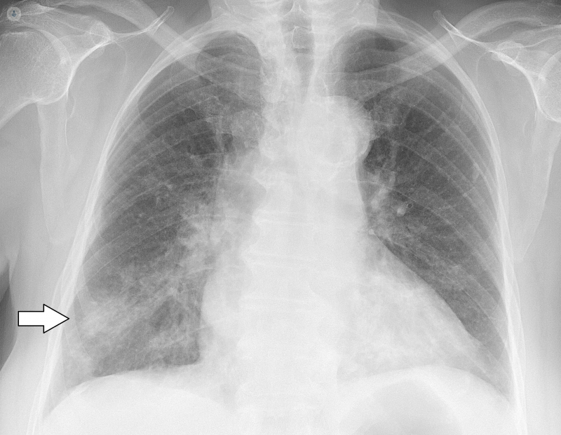
What does an abnormal chest x-ray look like?
Lungs: the whiter the lungs appear on the scan, the more dense they will be; the darker they are, the less dense. A doctor will assess which side of the lungs or if both sides are abnormal based on key surrounding features.
Heart: the heart blocks radiation so will appear lightly in the scan. Any abnormalities with the heart or surrounding it will appear as lighter or darker areas.
Bones: bones are the densest natural structure seen in a chest x-ray. Fractures will be quickly and easily seen in the scan as dark lines through the bone.
A radiologist will look for clues indicating there is an abnormality and will report with the doctor who ordered the chest x-ray.
What's the next step?
If the doctor is concerned by an abnormal chest X-ray, but doesn’t have enough information to make a diagnosis, they may order further tests, including a chest CT scan or a PET scan. By analysing the results of all these tests, the problem can be identified, and the doctor can plan a course of action with the patient to treat the condition.
- Most viewed diseases, medical tests, and treatments
- DEXA scan
- Ulnar nerve surgery
- Back pain
- Long Covid
- Anti-ageing treatments
- Digestive diseases
- Medicolegal
- Vertigo
- Anxiety
- Genetic study of Fragile X syndrome