Echocardiogram – Part 3: Types of heart scan
Escrito por:How does a doctor look inside your chest at your heart? How can a cardiologist check that all the chambers, blood vessels and valves are working properly? How are heart problems found? Top cardiologist Dr Allan Harkness concludes his series on echocardiograms with this guide to the different scans used to monitor heart health.
What are the more advanced echocardiograms I might need after a standard echocardiogram?
An echocardiogram is often one of the first tests done to look at a heart’s function. This is because it is completely safe, with no radiation or injections and it is a relatively quick and easy test.
However, sometimes the pictures are not as clear as we need or special techniques are required to get a better understanding of the heart. There are several more advanced echocardiograms we can use to help:

which is usually black on a normal echocardiogram.
Contrast echocardiogram
A special bubble contrast solution is injected into a vein in your arm and then the echocardiogram is done while the solution is inside your heart. This gives a clearer picture of the overall function of the main pumping chambers of your heart.
Bubble echocardiogram
A small amount of salt water with very tiny air bubbles is injected into a vein

tiny bubbles crossing from right to left
(the image is mirrored by convention).
in your arm and the echocardiogram done as the solution passes through your heart. This test is done to see if you might have a hole in your heart. You may be asked to do some odd breathing manoeuvres to help show up the hole.
Stress echocardiogram
This test looks to see how well your heart works when it is beating vigorously. If you have problems with the arteries of your heart or with the heart valves, this might only show up when the heart is beating fast. A stress echo involves taking pictures of your heart at rest and when it is beating fast. To make it go faster, we can either get you to exercise or else inject medication in the vein in your arm which speeds it up.

inside the heart (this is exceptionally rare)
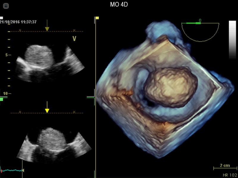
Transoesophageal echocardiogram
Your gullet (oesophagus) runs down behind your heart. A thin ultrasound probe passed down your gullet can look at your heart from behind and give exceptionally clear pictures.
This is particularly useful for looking at damaged heart valves and is often done for patients who may require an operation on their heart valves. The 3D pictures we can get from this form of echocardiogram are close to what the surgeon sees when they cut open your heart and peer inside.
This test can also look for holes in the heart or any signs of clots that may cause a stroke.
Are any other tests needed to confirm results of an echocardiogram?
If the standard or advanced echocardiograms do not give all the information required, you might need other tests to look at your heart.
Cardiac MRI
This form of scan uses powerful magnets to look at the heart. No radiation is used but you will need an injection of special contrast in a vein in your arm to help with the pictures. It will involve you lying in a tunnel for some time which may be a problem if you are very claustrophobic.
Cardiac MRI gives incredible detailed pictures of your heart and can complement the information seen on an echocardiogram. It is particularly useful for looking at the texture of the muscle of the heart.
Cardiac MRI is often used to see why a heart is not working well as it can show up inflammation or scarring. Cardiac MRI is also very accurate at telling how badly a heart valve is failing.
Although every hospital has an MRI machine, cardiac MRI is a very specialist technique and not every hospital has a dedicated machine or doctors skilled to perform it. This means you may need to travel to a specialist centre for a cardiac MRI.

Cardiac CT
Modern CT scanners can take very detailed pictures of the heart and the latest machines can do this with very little radiation. The main use of cardiac CT is to look at the heart’s coronary arteries.
If your symptoms suggest you might have angina or your ECG or echocardiogram showed signs that you may have furring up of your coronary arteries, then a Cardiac CT can help.
CT coronary angiography is now the initial test NICE recommends for people complaining of chest pain who might have angina. It is very good at ruling out coronary disease. When it does find narrowed arteries, your cardiologist will start you on protective medication. You might need further tests to look at the arteries in even more detail or to tell how much of the heart is affected by the blockages.
Invasive coronary angiography

This is the traditional way to look at the heart’s arteries and involves passing a thin tube up the artery in your wrist to your heart. This is clearly more invasive than a CT scan but can give even clearer pictures of any narrowing or blockage. It may help decide if you need stents or bypass surgery to improve the blood flow to your heart.


