What is a cardiac MRI?
MRI scans are a non-invasive diagnostic test. MRI uses powerful magnets, radio frequency pulses and a computer to create detailed pictures of soft tissues, organs, bone and other bodily structures. A cardiac MRI therefore produces images of the heart and surrounding tissue. Unlike X-ray, MRI does not use radiation.
What does cardiac MRI consist of?
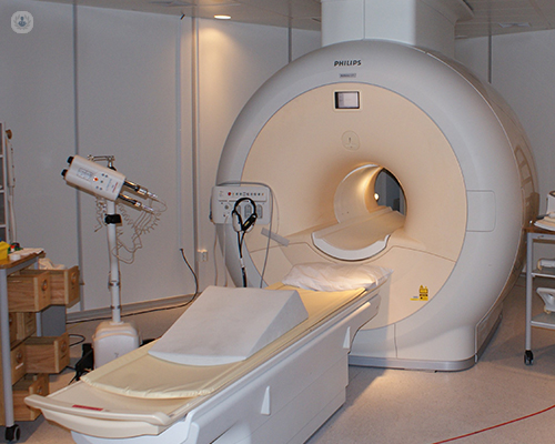
An MRI machine looks like a large tube with a flat bench that glides in and out of the tube. During an MRI you lie on the bench. The technician stays in a separate room during the scan, communicating with a microphone. When the scan begins, there can be a loud whirring noise as the machine takes pictures. You may also be asked to hold your breath at certain points by the technician to aid in getting better pictures. Sometimes contrast material will be used to highlight certain areas of the heart. This would be injected into the bloodstream. Beforehand, the technician will ask you about any possible allergies.
Why is cardiac MRI done?
A cardiac MRI is used to detect and monitor cardiac disease, including:
- Diagnosing cardiovascular disease
- Checking for infections, tumours and inflammation
- Checking the effects of coronary artery disease and the blood flow within the heart
- Monitoring congenital heart conditions
- Detecting heart valve defects
- Evaluating damage from a heart attack
How do you preparation for a cardiac MRI?
If you have a pacemaker, it's important to inform your specialist. If you do have a pacemaker, it might be necessary to have a CT scan instead, as an MRI can sometimes disrupt certain types of pacemakers. It's also important to inform your doctor whether or not you have any of the following (this is because the MRI machine uses strong magnets):
- Pins
- Staples
- Stents
- Artificial valves
- Implants
- Screws
How does it feel during a cardiac MRI?
A heart MRI is painless and non-invasive. They can take up to 90 minutes, depending on the purpose of the scan. It can sometimes be cold in the imaging room, so a blanket can be provided. It is also important that the patient remains still during the scan, which can be difficult for longer periods. The machine can be quite loud, so sometimes ear plugs can be provided.
What are the risks of a cardiac MRI?
Magnetic resonance does not use any radiation and is very safe with few, if any side-effects. The type of contrast that is often used is gadolinium, which is very safe and rarely causes any type of allergic reaction. However, the strong magnetic fields created during the test can cause some implants such as pacemakers not to work as well. Magnets can also cause a piece of metal inside the body to move or change position, but this should not happen with careful preparation.
11-13-2012 10-06-2023Cardiac MRI
Dr Rajesh Chelliah - Cardiology
Created on: 11-13-2012
Updated on: 10-06-2023
Edited by: Karolyn Judge
What is a cardiac MRI?
MRI scans are a non-invasive diagnostic test. MRI uses powerful magnets, radio frequency pulses and a computer to create detailed pictures of soft tissues, organs, bone and other bodily structures. A cardiac MRI therefore produces images of the heart and surrounding tissue. Unlike X-ray, MRI does not use radiation.

What does cardiac MRI consist of?
An MRI machine looks like a large tube with a flat bench that glides in and out of the tube. During an MRI you lie on the bench. The technician stays in a separate room during the scan, communicating with a microphone. When the scan begins, there can be a loud whirring noise as the machine takes pictures. You may also be asked to hold your breath at certain points by the technician to aid in getting better pictures. Sometimes contrast material will be used to highlight certain areas of the heart. This would be injected into the bloodstream. Beforehand, the technician will ask you about any possible allergies.
Why is cardiac MRI done?
A cardiac MRI is used to detect and monitor cardiac disease, including:
- Diagnosing cardiovascular disease
- Checking for infections, tumours and inflammation
- Checking the effects of coronary artery disease and the blood flow within the heart
- Monitoring congenital heart conditions
- Detecting heart valve defects
- Evaluating damage from a heart attack
How do you preparation for a cardiac MRI?
If you have a pacemaker, it's important to inform your specialist. If you do have a pacemaker, it might be necessary to have a CT scan instead, as an MRI can sometimes disrupt certain types of pacemakers. It's also important to inform your doctor whether or not you have any of the following (this is because the MRI machine uses strong magnets):
- Pins
- Staples
- Stents
- Artificial valves
- Implants
- Screws
How does it feel during a cardiac MRI?
A heart MRI is painless and non-invasive. They can take up to 90 minutes, depending on the purpose of the scan. It can sometimes be cold in the imaging room, so a blanket can be provided. It is also important that the patient remains still during the scan, which can be difficult for longer periods. The machine can be quite loud, so sometimes ear plugs can be provided.
What are the risks of a cardiac MRI?
Magnetic resonance does not use any radiation and is very safe with few, if any side-effects. The type of contrast that is often used is gadolinium, which is very safe and rarely causes any type of allergic reaction. However, the strong magnetic fields created during the test can cause some implants such as pacemakers not to work as well. Magnets can also cause a piece of metal inside the body to move or change position, but this should not happen with careful preparation.

What are the different kinds of non-invasive cardiac imaging tests?
By Dr Yousef Daryani
2024-11-21
You may have been told by your doctor that you need cardiac imaging to check for heart disease, or you may have symptoms of heart disease and want to be checked by a specialist. With the help of leading consultant cardiologist, Dr Yousef Daryani talks through the different procedures, and what to expect. See more


What are the advantages and disadvantages of cardiac MRI scans?
By Dr Thomas Heseltine
2024-11-21
Cardiac MRI (magnetic resonance imaging) is a non-invasive imaging technique used to assess the structure and function of the heart. It provides detailed images of the heart and surrounding blood vessels, helping to diagnose various cardiac conditions. Like all medical procedures, cardiac MRI scans come with both advantages and disadvantages. Here to tell us all about is leading consultant cardiologist Dr Thomas Heseltine. See more

How to investigate palpitations
By Dr Malcolm Finlay
2024-11-17
Experiencing palpitations can be a cause for concern, and you may be wondering how they're diagnosed. Leading consultant cardiologist and electrophysiologist Dr Malcolm Finlay goes into expert detail to explain how palpitations are medically investigated, in this informative article. See more


Heart failure: Your guide to diagnosis, treatment and helpful lifestyle modifications
By Dr Teresa Castiello
2024-11-11
Revered consultant cardiologist Dr Teresa Castiello explains how heart failure is diagnosed and treated in this article. In addition, the leading specialist shares specialist insight on the most beneficial lifestyle modifications patient can make. See more
Experts in Cardiac MRI
-
Dr Aigul Baltabaeva
CardiologyExpert in:
- Valvular heart disease
- Hypertension (high blood pressure)
- Chest pain
- Echocardiogram
- Cardiac MRI
- Blood pressure test
-
Dr Sabiha Gati
CardiologyExpert in:
- Sports cardiology
- Cardiomyopathy
- Cardiac screening
- Cardiac MRI
- Echocardiogram
- Stress test
-
Professor Mirza Kamran Baig
CardiologyExpert in:
- Interventional cardiology
- Heart failure
- Cardiomyopathy
- Arrhythmia
- Cardiac MRI
- Shortness of breath
-
Dr Thomas Heseltine
CardiologyExpert in:
- Cardiac MRI
- Coronary CT
- Imaging diagnostic system
- Heart failure
- Hypertension (high blood pressure)
- Chest pain
-
Dr Jonathan Hasleton
CardiologyExpert in:
- Chest pain
- Angina
- Shortness of breath
- Coronary heart disease
- Interventional cardiology
- Cardiac MRI
- See all

Goring Hall Hospital - part of Circle Health Group
Goring Hall Hospital - part of Circle Health Group
Bodiam Ave, Goring-by-Sea, Worthing BN12 5AT
No existe teléfono en el centro.
By using the telephone number provided by TOP DOCTORS, you automatically agree to let us use your phone number for statistical and commercial purposes. For further information, read our Privacy Policy
Top Doctors

Chiswick Medical Centre (HCA)
Chiswick Medical Centre (HCA)
347 - 353 Chiswick High Road
No existe teléfono en el centro.
By using the telephone number provided by TOP DOCTORS, you automatically agree to let us use your phone number for statistical and commercial purposes. For further information, read our Privacy Policy
Top Doctors

London Bridge Hospital - part of HCA Healthcare
London Bridge Hospital - part of HCA Healthcare
27 Tooley St
No existe teléfono en el centro.
By using the telephone number provided by TOP DOCTORS, you automatically agree to let us use your phone number for statistical and commercial purposes. For further information, read our Privacy Policy
Top Doctors
-
Goring Hall Hospital - part of Circle Health Group
Bodiam Ave, Goring-by-Sea, Worthing BN12 5AT, Goring-by-SeaExpert in:
- Hip
- Cataracts
- Gallbladder surgery
- General Surgery
- Orthopaedic surgery
- Hernia
-
Chiswick Medical Centre (HCA)
347 - 353 Chiswick High Road, West LondonExpert in:
- Dermatology
- Diagnostic Imaging
- Obstetrics and Gynaecology
- Sports Medicine
- General practice
-
London Bridge Hospital - part of HCA Healthcare
27 Tooley St, Central LondonExpert in:
- 24-hour service
- Cardiology
- Minimal access surgery (keyhole surgery)
- Orthopaedic surgery
- Cardiovascular disease
- Gastroenterology
- Most viewed diseases, medical tests, and treatments
- Migraine
- Paediatric rheumatology
- Autoimmune diseases
- Joint pain
- Child nutrition
- Nutrition
- Genetic testing
- Testicular ultrasound
- Breast ultrasound
- Abdominal pain


