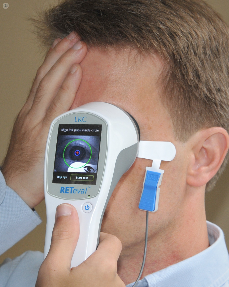
Electroretinography
Dr Taimour Alam - Neurophysiology
Created on: 07-12-2013
Updated on: 07-03-2023
Edited by: Sophie Kennedy
What is electroretinography?
Electroretinography, also known as electroretinogram, is a diagnostic test used to measure the electrical response of the cells of the retina (the back wall of the eye) when exposed to light.

The rod and cone cells that make up the retina are light-sensitive and are essential to our ability to see. They react to light and cause an electrical signal that is sent via the adjoining ganglion cells through the optic nerve to the brain. There, the brain interprets the signal as visual information, and this is how we see what we see. There are roughly 120 million rods and between 6 and 7 million cones in the human eye.
An electroretinogram examines the rods and cones and their connecting ganglions to check that visual information sent from these cells is reaching the brain correctly. Electroretinogram testing is performed by specialists in ophthalmology to diagnose various conditions which affect the retina.
What does electroretinography testing involve?
Electroretinography involves placing an electrode on the cornea (the front of the eye) in order to measure the electrical signals, and then presenting the eye with stimuli, such as flashes of light and reversing chequered patterns.
Why is electroretinography testing performed?
An ERG can be used for diagnosing a number of retinal conditions, such as:
- Retinitis pigmentosa
- Macular degeneration
- Retinoblastoma (a cancer of the retina)
- Retinal detachment
- Cone rod dystrophy (CRD)
It can also be useful in assessing if ocular surgery is required.
Preparation for electroretinography
The patient’s eyes must be dilated first with normal dilating eye drops. Then the eyes are numbed with anaesthetic drops. The patient is put in a comfortable position, either lying or sitting.
What to expect during the test
Once the patient is ready, with their eyes numbed by the anaesthetic eye drops, a speculum is used to hold their eyelids open. Then an electrode is gently placed onto each eye via what resembles a contact lens and an additional electrode is placed on the skin.
The patient then watches light stimuli, such as flashes, and the electrodes monitor the response of the retina to the light.
