What is the retina?
The retina is a thin layer of tissue on the back wall of the eye made up of different cells (retinal pigment epithelium, nerve cells and photoreceptor cells). The photoreceptor cells (cones and rods) react to light, and send electrical signals to the brain, which interprets these signals as vision.
The retina, along with the optic nerve, develops in the embryo as part of the brain tissue, making it part of the central nervous system. Being a nervous tissue, the retina doesn’t have the ability to regenerate itself.
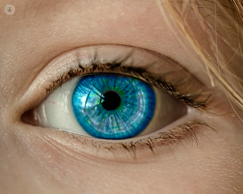
What does the retina do?
The retina is responsible for our vision. The photoreceptor cells (cones and rods) react to light entering the eye, releasing chemicals, which in turn trigger a nerve impulse, which is sent to the cerebral cortex via the optic nerve. The pattern of light hitting the photoreceptor cells triggers a unique set of electrical signals, which are interpreted by the brain as an image.
Rod cells function well in low light, providing us with our night vision. They contribute little to colour vision, which is why we find it difficult to distinguish colours in darkness. They tend to be concentrated around the edge of the retina and are also used for peripheral vision.
Cone cells, on the other hand, are less sensitive to light, making them of little use in low-light conditions. However, they are responsible for our colour vision. Most people have three kinds of cone cell. Each type is sensitive to a different visible wavelengths of light, giving us trichromatic vision. The mixture of signals sent by the different cone cells being stimulated to different degrees creates the mixture of colours we can perceive. Some people are born with only two types of cone cells, limiting the range of colours they can perceive, causing them to easily confuse colours that appear different to someone with all three types of cone cell.
What conditions can affect it?
Retinal conditions can affect your eyesight with different degrees of severity. They can be either acquired or inherited:
- Diabetic retinopathy – diabetes causes the capillaries that supply the retina to leak fluid into or under the retina, leading to swelling and distorted vision.
- Retinal tear – when the gel-like vitreous inside the eye shrinks it can tug at the retina, and sometimes this can cause a tear.
- Retinal detachment – a disorder in which the retina becomes loose and detaches from the tissue underneath. It is usually caused when fluid passes through a retinal tear.
- Retinitis pigmentosa – a degenerative disease of the retina
- Macular degeneration (AMD) – age-related deterioration of the macula – a small part of the retina with a high concentration of cone cells, responsible for central colour vision.
- Macular hole – a small defect in the macula, perhaps caused by trauma or traction between the retina and the vitreous.
- Macular oedema - swelling in the retina caused by a number of conditions. Macular oedema is very common in uveitis and is often a driver for treatment.
How can such conditions be treated?
The treatment will vary according to the specific condition. Some conditions can be treated with intravitreal injections; for others, the only option is surgery.
Which doctor should I see?
You should see a specialised ophthalmologist.
05-12-2017 09-13-2023Retina
Miss Rahila Zakir - Ophthalmology
Created on: 05-12-2017
Updated on: 09-13-2023
Edited by: Conor Lynch
What is the retina?
The retina is a thin layer of tissue on the back wall of the eye made up of different cells (retinal pigment epithelium, nerve cells and photoreceptor cells). The photoreceptor cells (cones and rods) react to light, and send electrical signals to the brain, which interprets these signals as vision.
The retina, along with the optic nerve, develops in the embryo as part of the brain tissue, making it part of the central nervous system. Being a nervous tissue, the retina doesn’t have the ability to regenerate itself.

What does the retina do?
The retina is responsible for our vision. The photoreceptor cells (cones and rods) react to light entering the eye, releasing chemicals, which in turn trigger a nerve impulse, which is sent to the cerebral cortex via the optic nerve. The pattern of light hitting the photoreceptor cells triggers a unique set of electrical signals, which are interpreted by the brain as an image.
Rod cells function well in low light, providing us with our night vision. They contribute little to colour vision, which is why we find it difficult to distinguish colours in darkness. They tend to be concentrated around the edge of the retina and are also used for peripheral vision.
Cone cells, on the other hand, are less sensitive to light, making them of little use in low-light conditions. However, they are responsible for our colour vision. Most people have three kinds of cone cell. Each type is sensitive to a different visible wavelengths of light, giving us trichromatic vision. The mixture of signals sent by the different cone cells being stimulated to different degrees creates the mixture of colours we can perceive. Some people are born with only two types of cone cells, limiting the range of colours they can perceive, causing them to easily confuse colours that appear different to someone with all three types of cone cell.
What conditions can affect it?
Retinal conditions can affect your eyesight with different degrees of severity. They can be either acquired or inherited:
- Diabetic retinopathy – diabetes causes the capillaries that supply the retina to leak fluid into or under the retina, leading to swelling and distorted vision.
- Retinal tear – when the gel-like vitreous inside the eye shrinks it can tug at the retina, and sometimes this can cause a tear.
- Retinal detachment – a disorder in which the retina becomes loose and detaches from the tissue underneath. It is usually caused when fluid passes through a retinal tear.
- Retinitis pigmentosa – a degenerative disease of the retina
- Macular degeneration (AMD) – age-related deterioration of the macula – a small part of the retina with a high concentration of cone cells, responsible for central colour vision.
- Macular hole – a small defect in the macula, perhaps caused by trauma or traction between the retina and the vitreous.
- Macular oedema - swelling in the retina caused by a number of conditions. Macular oedema is very common in uveitis and is often a driver for treatment.
How can such conditions be treated?
The treatment will vary according to the specific condition. Some conditions can be treated with intravitreal injections; for others, the only option is surgery.
Which doctor should I see?
You should see a specialised ophthalmologist.

Vitrectomy surgery: What to expect and what are the risks?
By Mr Danny Mitry
2024-11-21
If you have retinal or vitreous problems, you may require vitrectomy, or vitreoretinal surgery. It's an operation that can treat these issues. Leading ophthalmologist Mr Danny Mitry goes into expert detail about what to expect and the risks of vitrectomy surgery in this informative article. See more

Your guide to full-thickness macular holes and surgery
By Mr Alistair Laidlaw
2024-11-20
When a gap forms all the way through the retina, it's referred to as a full-thickness macular hole. This can cause vision to become darker and blurry but fortunately, there is surgical treatment available to close a macular hole and therefore prevent further deterioration of vision. Mr Alistair Laidlaw provides a comprehensive guide that clarifies symptoms, treatment, the success rate of treatment and more. See more

Comprehensive guide to full-thickness macular hole surgery and care
By Professor James Bainbridge
2024-11-20
Macular holes can lead to distorted vision and blind spots. If you have or think you may have a macular hole, surgery provides a promising chance of repairing it and preserving your eyesight. Professor James Bainbridge provides you with an in-depth understanding of macular hole surgery and aftercare. See more


Retinitis pigmentosa: the night blindness disease
By Mr Niaz Islam
2024-11-20
Retinitis pigmentosa is an inherited retinal dystrophy and it affects the light-sensitive cells of the retina. Mr Niaz Islam explains just how retinitis pigmentosa affects vision, whether it can lead to complete blindness and if there’s hope for finding a cure. See more
Experts in Retina
-
Mr Niaz Islam
OphthalmologyExpert in:
- Cataracts
- Chalazion
- Diabetic retinopathy
- Macular degeneration (AMD)
- Retina
- Watery eyes
-
Mr Praveen Patel
OphthalmologyExpert in:
- Macular degeneration (AMD)
- Retina
- Retinal vein occlusion
- Diabetic retinopathy
- Macular oedema
- Intravitreal injection
-
Professor Adnan Tufail
OphthalmologyExpert in:
- Retina
- Macular degeneration (AMD)
- Diabetic retinopathy
- Cataracts
- Myopia
- Retinal vein occlusion
-
Mr Roger Wong
OphthalmologyExpert in:
- Retina
- Diabetic retinopathy
- Retinal detachment
- Macular hole
- Epiretinal membrane
- Macular degeneration (AMD)
-
Mr Ulrich Meyer-Bothling
OphthalmologyExpert in:
- Cataracts
- Lens replacement (intraocular lenses)
- Glaucoma
- Retina
- Diabetic retinopathy
- YAG laser capsulotomy
- See all

Optegra Yorkshire Eye Hospital
Optegra Yorkshire Eye Hospital
Optegra Eye Hospital Yorkshire, 937 Harrogate Road, Apperley Bridge, Bradford BD10 0RD
No existe teléfono en el centro.
By using the telephone number provided by TOP DOCTORS, you automatically agree to let us use your phone number for statistical and commercial purposes. For further information, read our Privacy Policy
Top Doctors

Laser Vision
Laser Vision
Prema Compass Road North Harbour Business Park Portsmouth PO6 4RP
No existe teléfono en el centro.
By using the telephone number provided by TOP DOCTORS, you automatically agree to let us use your phone number for statistical and commercial purposes. For further information, read our Privacy Policy
Top Doctors

Great Ormond Street Hospital
Great Ormond Street Hospital
Great Ormond Street. WC1N 3JH
No existe teléfono en el centro.
By using the telephone number provided by TOP DOCTORS, you automatically agree to let us use your phone number for statistical and commercial purposes. For further information, read our Privacy Policy
Top Doctors
-
Optegra Yorkshire Eye Hospital
Optegra Eye Hospital Yorkshire, 937 Harrogate Road, Apperley Bridge, Bradford BD10 0RD, BradfordExpert in:
- Cataracts
- Laser eye surgery
- Refractive surgery
- ICL lens implants
- Lens replacement
-
Laser Vision
Prema Compass Road North Harbour Business Park Portsmouth PO6 4RP, HavantExpert in:
- Cataracts
- Laser eye surgery
- ICL lens implants
- Ophthalmology
- Keratoconus
- Lens replacement
-
Great Ormond Street Hospital
Great Ormond Street. WC1N 3JH, Central LondonExpert in:
- Cancer
- Paediatric neurosurgery
- Paediatrics
- See all
- Most viewed diseases, medical tests, and treatments
- Genetic testing
- Minimal access surgery (keyhole surgery)
- Botulinum toxin (Botox™)
- Orthoptics
- Medicolegal
- Dermal fillers
- Headache
- Strabismus (squint)
- Glaucoma
- Diplopia (double vision)


