What is an angiography?
An angiography is a type of X-ray which examines the blood vessels, particularly the arteries, veins and heart chambers.
What happens during an angiography?
First, a catheter is inserted into one of your arteries, depending on the area which needs to be examined. Through the catheter, a dye is administered to contrast the area that needs to be visualised. The catheter is then guided through the arterial system to the area which needs to be examined. X-rays are then taken as the contrast dye flows through the blood vessels.
Why is an angiography performed?
An angiography is done to check how the blood flows through the vessels, and the general health of the blood vessels too. An angiography is useful in investigating and diagnosing problems affecting the blood vessels, such as narrowing of the arteries, blood clots, or embolisms.
Preparing for angiography
Before the test, the patient is examined, including a blood test. The clinic or doctor will ask you about your medical history and if you are taking any medications. The doctor will also ask if you would prefer to be sedated during the procedure.
What does the exam feel like?
The X-ray table can be somewhat uncomfortable, and the procedure generally lasts between thirty minutes and two hours. You may also feel a pinch when local anaesthesia is administered, but the area is generally numbed so it shouldn’t hurt. When the catheter is inserted and moved around the body, you may feel a pushing or pulling sensation, but it should not be painful.
What happens after the test?
After the angiography, you will be moved to an area where you can rest, and you have to lie still for a few hours to prevent bleeding. Generally angiographies are performed as a day case procedure, although in some cases you may be kept in overnight for monitoring.
Generally results of the test are available after a few weeks. You may feel some discomfort, such as soreness at the entry site for a few days after the procedure. Some bruising may also occur.
Angiography
Dr Navin Chandra - Cardiology
Created on: 04-23-2015
Updated on: 09-15-2023
Edited by: Sophie Kennedy
What is an angiography?
An angiography is a type of X-ray which examines the blood vessels, particularly the arteries, veins and heart chambers.

What happens during an angiography?
First, a catheter is inserted into one of your arteries, depending on the area which needs to be examined. Through the catheter, a dye is administered to contrast the area that needs to be visualised. The catheter is then guided through the arterial system to the area which needs to be examined. X-rays are then taken as the contrast dye flows through the blood vessels.
Why is an angiography performed?
An angiography is done to check how the blood flows through the vessels, and the general health of the blood vessels too. An angiography is useful in investigating and diagnosing problems affecting the blood vessels, such as narrowing of the arteries, blood clots, or embolisms.
Preparing for angiography
Before the test, the patient is examined, including a blood test. The clinic or doctor will ask you about your medical history and if you are taking any medications. The doctor will also ask if you would prefer to be sedated during the procedure.
What does the exam feel like?
The X-ray table can be somewhat uncomfortable, and the procedure generally lasts between thirty minutes and two hours. You may also feel a pinch when local anaesthesia is administered, but the area is generally numbed so it shouldn’t hurt. When the catheter is inserted and moved around the body, you may feel a pushing or pulling sensation, but it should not be painful.
What happens after the test?
After the angiography, you will be moved to an area where you can rest, and you have to lie still for a few hours to prevent bleeding. Generally angiographies are performed as a day case procedure, although in some cases you may be kept in overnight for monitoring.
Generally results of the test are available after a few weeks. You may feel some discomfort, such as soreness at the entry site for a few days after the procedure. Some bruising may also occur.
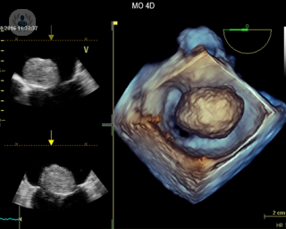

Echocardiogram – Part 3: Types of heart scan
By Dr Allan Harkness
2024-12-17
How does a doctor look inside your chest at your heart? How can a cardiologist check that all the chambers, blood vessels and valves are working properly? How are heart problems found? Top cardiologist Dr Allan Harkness concludes his series on echocardiograms with this guide to the different scans used to monitor heart health. See more
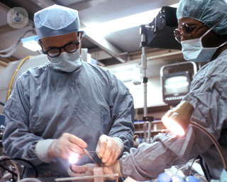

Exploring transcatheter aortic valve implantation (TAVI)
By Dr Mohamed Farag
2024-12-16
Transcatheter aortic valve implantation (TAVI), or TAVR, is a minimally invasive procedure used to treat aortic valve stenosis, a condition in which the aortic valve becomes narrowed, restricting blood flow from the heart to the rest of the body. Leading consultant interventional cardiologist Dr Mohamed Farag takes an in-depth look at TAVI and addresses some common questions patients may have about this procedure, in this article. See more

CT coronary angiography: How is it performed and what does it detect?
By Dr Aftab Gill
2024-12-15
A CT coronary angiography is a test that analyses the heart and the coronary arteries surrounding the heart to identify if there are any blockages or any other heart-related problems that need to be treated. In this article, expert consultant cardiologist Dr Aftab Gill explains in detail what exactly a CT coronary angiography is, how it's performed and what it detects. See more
Experts in Angiography
-
Mr Jonathan Dowler
OphthalmologyExpert in:
- Intravitreal injection
- Retinal tear
- Angiography
- Epiretinal membrane
- Macular degeneration (AMD)
- Diabetic retinopathy
-
Dr Nadeem Qazi
Interventional radiologyExpert in:
- Angiography
- Chemoembolization
- Fibroids
- Interventional oncology
- Radiofrequency ablation (RFA)
-
Dr Stuart Coley
RadiologyExpert in:
- Gamma knife
- Imaging diagnostic system
- Neuroradiology
- Vascular malformations
- Angiography
- Aneurysms embolization
-
Dr Arun Sebastian
RadiologyExpert in:
- Interventional oncology
- CT scan (CAT)
- MRI
- Biopsy
- Prostate artery embolisation
- Angiography
-
Dr Brian Clapp
CardiologyExpert in:
- Angiography
- Coronary heart disease
- Atrial septal defect
- Patent foramen ovale
- Interventional cardiology
- Heart attack
- See all

Alliance Medical Marylebone
Alliance Medical Marylebone
10-11 Bulstrode Place, London. W1U 2HX
No existe teléfono en el centro.
By using the telephone number provided by TOP DOCTORS, you automatically agree to let us use your phone number for statistical and commercial purposes. For further information, read our Privacy Policy
Top Doctors

Amethyst: Thornbury Radiosurgery Centre
Amethyst: Thornbury Radiosurgery Centre
Thornbury Radiosurgery Centre, 312 Fulwood Rd, Sheffield, S10 3BR
No existe teléfono en el centro.
By using the telephone number provided by TOP DOCTORS, you automatically agree to let us use your phone number for statistical and commercial purposes. For further information, read our Privacy Policy
Top Doctors

London Medical
London Medical
49 Marylebone High Street, W1U 5HJ
No existe teléfono en el centro.
By using the telephone number provided by TOP DOCTORS, you automatically agree to let us use your phone number for statistical and commercial purposes. For further information, read our Privacy Policy
Top Doctors
-
Alliance Medical Marylebone
10-11 Bulstrode Place, London. W1U 2HX, Central LondonExpert in:
- Cardiology
- Diagnostic Imaging
- Ultrasound
- Gastroenterology
- Neurology
- Magnetic resonance
-
Amethyst: Thornbury Radiosurgery Centre
Thornbury Radiosurgery Centre, 312 Fulwood Rd, Sheffield, S10 3BR, SheffieldExpert in:
- Vascular Surgery
- Neurosurgery
- Neurology
- Medical Oncology
- Cancer Treatment
- Brain and spinal tumours
-
London Medical
49 Marylebone High Street, W1U 5HJ, Central LondonExpert in:
- Cardiology
- Adult Diabetes
- Child Diabetes
- Endocrinology
- General practice
- Ophthalmology
- See all
- Most viewed diseases, medical tests, and treatments
- Alzheimer's disease
- Electrophysiology study
- Visual impairment
- Diabetic retinopathy
- Retina
- Presbyopia
- Nystagmus
- Myopia
- Hyperopia (farsightedness)
- Eye examination


