Retinal detachment surgery

Mr Sidath Liyanage - Ophthalmology
Created on: 03-04-2013
Updated on: 03-24-2023
Edited by: Jay Staniland
What is retinal detachment surgery?
The retina is the light-sensitive layer of tissue that lines the inside of the eye and sends visual messages via the optic nerve to the brain. When the retina detaches, it is pulled from its normal position. Retinal detachment is caused by a retinal tear and can lead to blurred or lost vision.
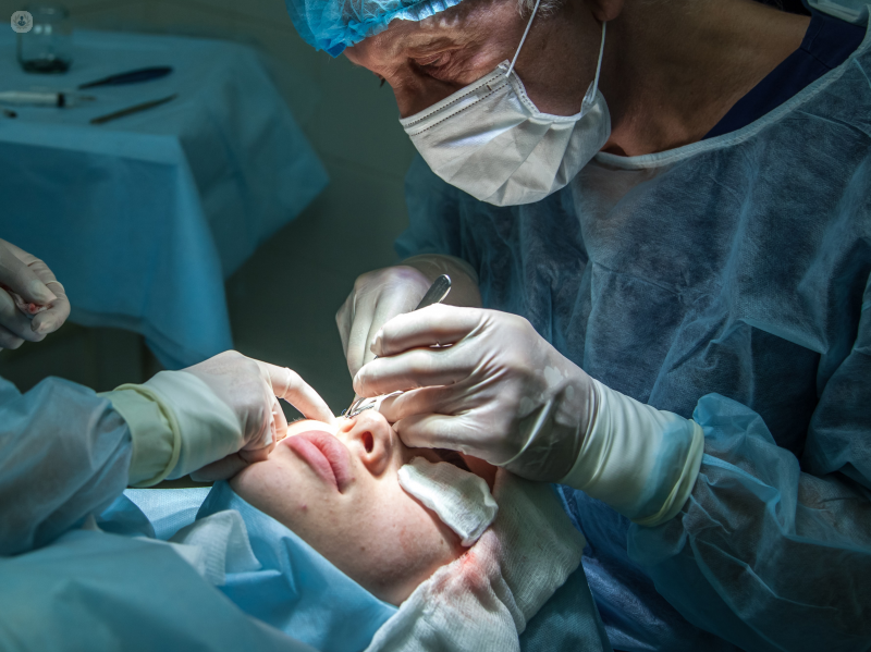
Surgery is the only treatment for retinal detachment. The goals of retinal detachment surgery are to reattach the retina and to prevent vision loss.
What is retinal detachment?
Retinal detachment is an eye condition that involves the separation of the retina from the underlying tissues within the eye. Retinal detachments are usually caused by a retinal break, hole or tear. These types of detachments are known as rhegmatogenous retinal detachments and occur when the vitreous gel (a clear liquid that fills two-thirds of the inside of the eye) pulls loose or separates from its attachment to the retina.
When the retina has torn, the vitreous gel passes through the tear and gathers behind the retina. This build-up behind the retina is what separates the retina from the choroid and retinal pigment epithelium (lining tissue). As more liquid vitreous collects behind the retina, it can lead to a total retinal detachment. It usually affects one eye at a time.
Types of procedures for retinal detachment surgery:
There are three types of eye surgery to treat retinal detachment, which are performed by an ophthalmologist.
- Vitrectomy – this is the most common surgical procedure for retinal detachment. The procedure removes the vitreous gel from the middle of the eye. The doctor uses small tools to remove the vitreous gel and replace it with either a gas or silicone bubble to hold the retina in position from the inside.
- Scleral buckling – this involves fine bands of silicone rubber or sponge that are stitched onto the outside of the white of the eye (the sclera), where the retina has detached. The bands act like a buckle, which presses the sclera in towards the middle of the eye so that the torn retina can lie against the wall of the eye. Laser or freezing treatment is used to scar the tissue around the retina, in order to create a seal between the retina and the wall of the eye.
- Pneumatic retinopexy – this procedure is often used if the detachment is small and uncomplicated. A small bubble of gas is injected into the eye to press the retina back into place. Laser or freezing treatment is often then used to create scar tissue to keep the retina in place. Over the next few weeks, the bubble is slowly absorbed into the eye.
Risks of retinal detachment surgery
There are a few complications, which are not too common but may occur following a surgical procedure and another operation may be needed to fix the retina. Possible complications include:
- Bleeding inside the eye
- More holes in the retina
- Bruises
- Glaucoma – swelling inside of the eye
- Cataracts – cloudy lens
- Double vision
- Infection

Everything you need to know about retinal detachment
By Mr Felipe Dhawahir-Scala
2025-02-02
Retinal detachment is a very serious condition that if left untreated can result in blindness. Find out how retinal detachment happens and what the warning signs are from our leading ophthalmologist, Mr Felipe Dhawahir-Scala. See more
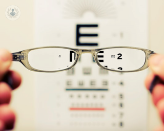

Myopia: An in-depth guide to short-sightedness
By Ms Evgenia (Jen) Anikina
2025-02-01
Myopia, or short-sightedness, can typically be corrected with glasses or contact lenses on prescription from an optician. The condition is increasingly common and isn't serious in itself but can lead to more serious complications. In this informative article, highly respected consultant vitreoretinal surgeon Ms Evgenia Anikina explains how myopia affects the eye and what can be done to slow its progress. See more


Retinal detachment surgery: Understanding the procedure
By Mr Palpandian Viswanathan
2025-02-01
Retinal detachment surgery is a procedure performed to repair a detached retina, a serious eye condition that requires prompt medical attention. The surgery aims to reattach the retina to the back of the eye, restoring vision and preventing further vision loss. In this comprehensive article, leading consultant ophthalmologist Mr Palpandian Viswanathan provides a detailed look at the procedure. See more


Retinal detachment repair using vitrectomy surgery
By Mr Vaughan Tanner
2025-01-31
A simple vitrectomy, performed because of vitreous floaters and debris, would have a 98-99% chance of a very successful outcome with no complications. Expert ophthalmologist Mr Vaughan Tanner explains more... See more
Experts in Retinal detachment surgery
-

Professor James Bainbridge
OphthalmologyExpert in:
- Cataracts
- Epiretinal membrane
- Macular hole
- Retinal detachment
- Eye floaters
- Retinal detachment surgery
-

Mr Robert Henderson
OphthalmologyExpert in:
- Retinal detachment surgery
- Paediatric ophthalmology
- Cataracts
- Cataracts in children
- Macular hole
- Epiretinal membrane
-

Mr Alistair Laidlaw
OphthalmologyExpert in:
- Retinal detachment surgery
- Macular hole
- Epiretinal membrane
- Macular degeneration (AMD)
- Diabetic retinopathy
- Cataracts
-
Mr Danny Mitry
OphthalmologyExpert in:
- Retinal detachment surgery
- Cataracts
- Refractive lens exchange
- Diabetic retinopathy
- Macular degeneration (AMD)
- Macular hole
-
Mr Craig Goldsmith
OphthalmologyExpert in:
- Retinal detachment surgery
- Cataracts
- Vitrectomy
- Epiretinal membrane
- Macular degeneration (AMD)
- Macular hole
- See all

Eye Surgery Scotland
Eye Surgery Scotland
40 Colinton Road, EH10 5BT
No existe teléfono en el centro.
By using the telephone number provided by TOP DOCTORS, you automatically agree to let us use your phone number for statistical and commercial purposes. For further information, read our Privacy Policy
Top Doctors
The Royal Free Hospital
The Royal Free Hospital
Pond Street, Hampstead. NW3 2QG
No existe teléfono en el centro.
By using the telephone number provided by TOP DOCTORS, you automatically agree to let us use your phone number for statistical and commercial purposes. For further information, read our Privacy Policy
Top Doctors
Great Ormond Street Hospital
Great Ormond Street Hospital
Great Ormond Street. WC1N 3JH
No existe teléfono en el centro.
By using the telephone number provided by TOP DOCTORS, you automatically agree to let us use your phone number for statistical and commercial purposes. For further information, read our Privacy Policy
Top Doctors
-

Eye Surgery Scotland
40 Colinton Road, EH10 5BT, EdinburghExpert in:
- Cataracts
- Retina and Vitreous
- Lens replacement
-
The Royal Free Hospital
Pond Street, Hampstead. NW3 2QG, Central LondonExpert in:
- General Surgery
- Orthopaedic surgery
- Robotic Surgery
- Dermatology
- Obstetrics and Gynaecology
- Paediatrics
-
Great Ormond Street Hospital
Great Ormond Street. WC1N 3JH, Central LondonExpert in:
- Cancer
- Paediatric neurosurgery
- Paediatrics
- See all
- Most viewed diseases, medical tests, and treatments
- Visual impairment
- Diabetic retinopathy
- Retina
- Presbyopia
- Nystagmus
- Myopia
- Hyperopia (farsightedness)
- Eye examination
- Blepharitis
- Astigmatism